
Imaging
 Scanning electron microscope that can operate in high or low vacuum mode enabling the use of a wide variety of samples. Electron beam is generated by a tungsten filament which can resolve features down to 3 nanometers.
Scanning electron microscopy cathodoluminescence has become a key micro-analytical technique to image features within a variety of materials. The interaction of an electron beam with a sample generates multiple signals for micro-analysis, including secondary electrons related to surface morphology, backscattered electrons related to mean atomic number, characteristic X-rays related to elemental composition, and CL related to defects and trace element chemistry.
Example Use Cases:
- Measurement of samples at the nanometer level
- Quality control of nanofabrication (solar cells, computer chips)
Gatan Monarch Pro CL (Cathodoluminescence) System

We are dominantly using our CL system to image zircon mounts prior to geochronologic and geochemical analysis. Increasingly widespread applications of SEM-CL in the Earth Sciences include: imaging of zircon grains in igneous, metamorphic, and detrital sedimentary rocks to identify and target growth zones for geochronologic analyses; imaging of quartz to evaluate diagentic growth, alteration, and deformation microtextures;; and imagine of carbonate rocks to evaluate diagenetic, hydrothermal, and deformational components. Our multi-user facility is also utilizing our CL system to quantify impurities and defects in nanomaterials.
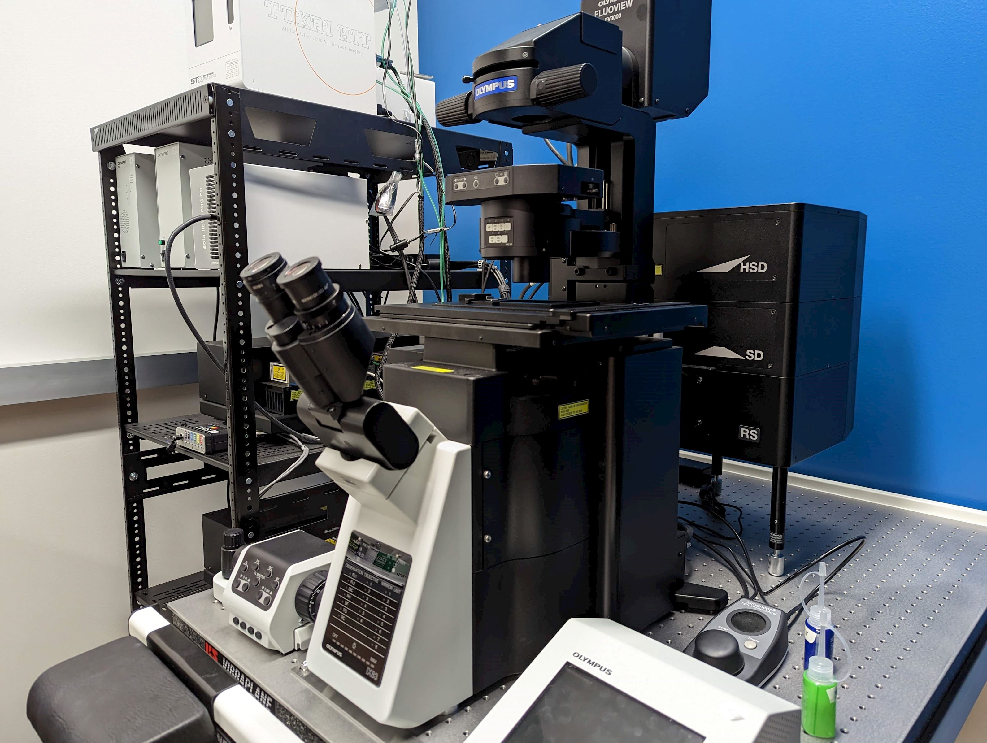 Featuring the high sensitivity and speed required for live cell imaging as well as deep tissue observation, the FV3000 confocal microscope enables a wide range of imaging modalities, including macro-to-micro imaging, super resolution microscopy, and quantitative data analysis.
Equipped with 5 different laser lights for visualizing different wavelengths. Samples can be stained with antibodies that are conjugated to chromophores that are then excited by the particular wavelength of laser light. This allows for different parts of a biological sample to be stained with different colors, Nucleus=Blue, DNA=Green, a particular protein=Red.
Example Use Cases:
- Imaging the inner structure of organelles
- Staining neurons to see the nucleus, synapse, dendrites, and axons
- Observing autofluorescence in microbiolates
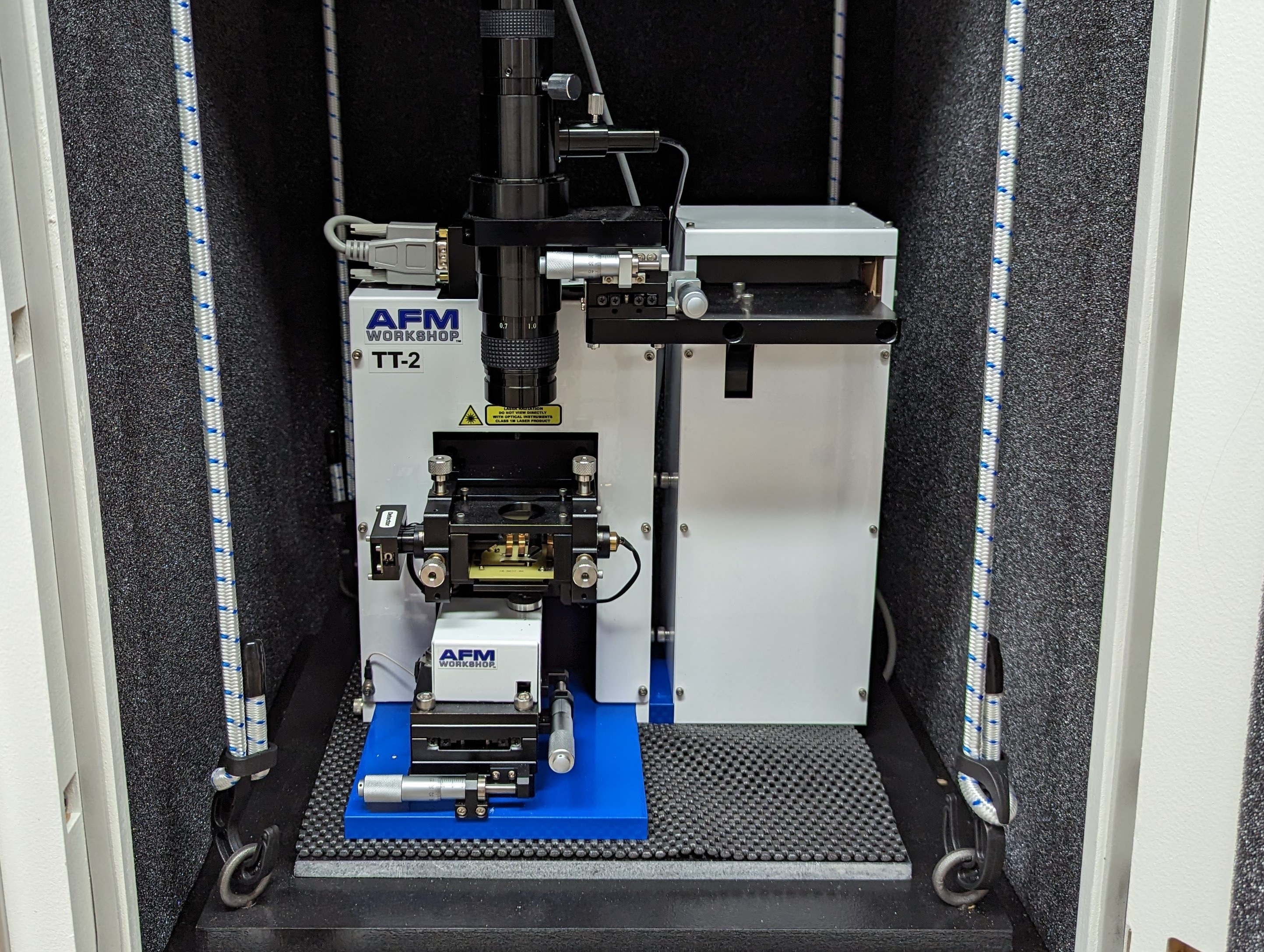
The Atomic Force Microscope (AFM) uses an ultra-sharp silicon crystal probe to scan over the surface of a sample. The tip is so sharp that it can sense the atomic forces along the surface of the sample to give precision down to hundreds of picometers (under a nanometer). This allows the instrument to give quantitative data on sample height.
The AFM is a complementary instrument to the Scanning Electron Microscope (SEM). While the SEM has the ability to look at samples with large differences in their heights the AFM excels at “flat” samples where the total height can be measured in nanometers. This allows the AFM to analyze samples in 3 dimensions as opposed to 2 dimensions in the SEM.
The AFM can measure samples without a vacuum, whereas the SEM need a vacuum to function.
Example Use Cases:
- Check fabrication of computer chips
- Imaging of cell membranes
- Viral morphology